Figure 3.

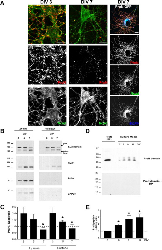
ProN is actively sorted to the cell surface during differentiation. A, Live hippocampal neurons (3 and 7 DIV) after staining with ProN antibody (red) were fixed and additionally stained for total Ncad (green). Note that the surface ProN levels diminish by 7 DIV and that ProN overexpression (cotransfection ProN/GFP, 4:1) leads to robust surface ProN expression. Scale bar, 10 μm. B, Biochemical evidence for surface sorting of ProN. Cell surface biotinylation supports the live staining data that ProN is actively sorted to the cell surface. The levels of surface (pulldowns) ProN drop significantly by 7 DIV, while the levels of mature N-cad increase markedly, coinciding with the elevation in the level of glutamate receptor (GluR1) at the cell surface. Actin and GADPH immunoblots show that the cytoplasmic proteins are spared from biotinylation. C, Quantification of ProN and N-cad levels in the total lysates and at the cell surface. The surface ProN/N-cad ratio drops significantly during differentiation. Results are expressed as mean values ± SD (n = 3 independent cultures). D, E, Release of processed prodomain by neurons. The ProN peptide accumulates in the culture media during neuronal differentiation. RP denotes the recombinant prodomain of ProN. The absence of bands when the immunoblot is incubated with ProN antibody that has been preincubation with RP demonstrates that the processed product in D is the prodomain of N-cad. Results are expressed as mean values ± SD (n = 3 independent cultures). *p < 0.001, compared with 3 DIV, by one-way ANOVA followed by Student–Newman–Keuls test.
