
Figure 6.
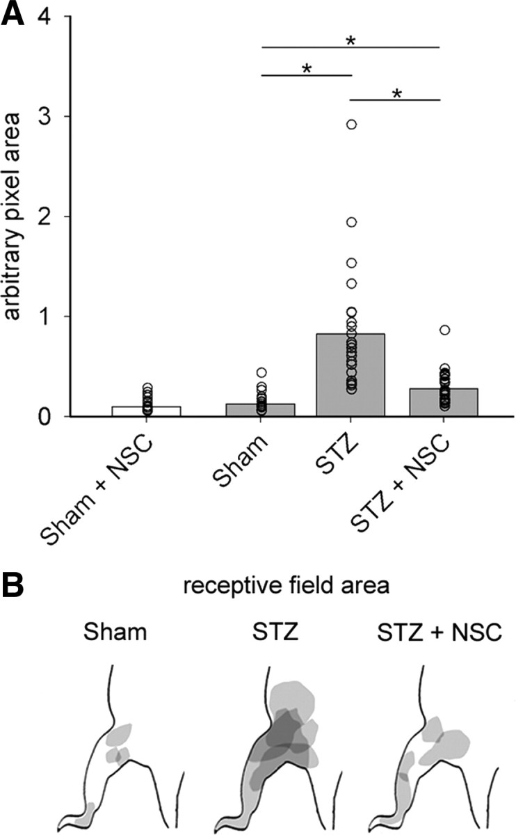
Receptive field areas expand in diabetic neuropathic pain. A, Quantification of WDR unit receptive field areas demonstrate that diabetic animals had receptive fields that were significantly larger, by more than sixfold, compared with Sham receptive field areas (one-way ANOVA with Bonferonni's post hoc, *p < 0.05). Treatment with NSC23766 decreased the size of receptive fields compared with untreated diabetic animals (one-way ANOVA with Bonferonni's post hoc, *p < 0.05), but not back to Sham levels (one-way ANOVA with Bonferonni's post hoc, *p < 0.05). NSC23766 treatment in Sham animals did not change receptive field size (white bar). B, A representative map shows that the receptive field areas in diabetic animals, STZ, increases compared with Sham and STZ animals treated with NSC23766, STZ plus NSC. In A, the open circles show individual animal means. The bar graph is mean ± SEM.