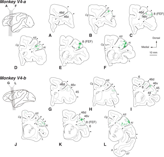
Figure 2.
Distribution patterns of retrograde labeling in the frontal cortex 3 d after the rabies injections into V4. Six representative coronal sections for monkeys V4-a and V4-b are arranged anteroposteriorly in A–F and G–L, respectively. For conventions, see Figure 1.

