Figure 1.

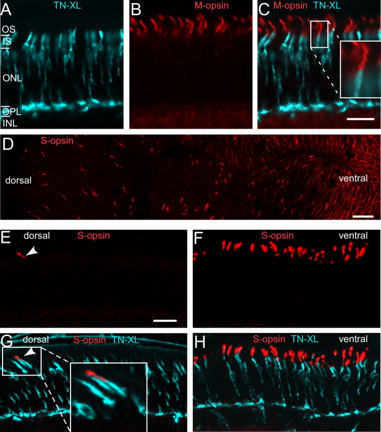
TN-XL expression in cone photoreceptors of the HR2.1:TN-XL mouse line. A–C, Vertical sections of HR2.1:TN-XL retina stained with antibodies against GFP (labels TN-XL; cyan) and M-opsin (red), showing the presence of the biosensor throughout the cone with the exception of the OS. Magnified inset in C illustrates that TN-XL and opsin labeling do not overlap. D, Retinal whole mount stained with antibodies against S-opsin, confirming the presence of the dorsal-ventral opsin coexpression gradient described in mouse (Szél et al., 1992). E–H, Vertical sections taken from the dorsal (E, G) and the ventral (F, H) retina and stained for S-opsin. Example for an S-opsin-expressing, TN-XL-positive cone in the dorsal retina (G, see also magnification in inset). ONL, Outer nuclear layer; INL, inner nuclear layer. Scale bars: A–C, E–H, 20 μm; D, 50 μm.
