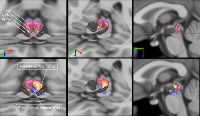
Figure 2.
MLR activations with respect to MLR structures in the basal ganglia atlas. Illustration of the MLR activations with respect to MLR nuclei [CN, dorsal PPN (PPNd), ventral PPN (PPNv), parts of the pedoculopontine nucleus) and the substantia nigra (SN). To better understand the overlap between the activations and the nuclei, we show three viewpoints of the nuclei (top) and the same viewpoints with the activations together with the nuclei (bottom). The orientation of the viewpoints is indicated by the arrows (red, right to left; green, back to front; blue, top to down). In the right column, the viewpoint is strictly sagittal, the horizontal line representing the transverse MRI section visible on the two other viewpoints at the level of the ventral PPN. The nuclei come from the 3D histological and deformable YeB atlas (Yelnik et al., 2007; Bardinet et al., 2009). This atlas has been mapped onto the MNI152 template through a validated intensity-based deformation procedure that is adapted to subcortical structures.

