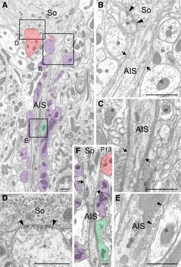
Figure 1.

Electron micrographs showing the pinceau formation and AIS of PC in adult (A–E) and postnatal day 13 (F) mice. A, F, Low-power images through the AIS. The AIS starts from the conical apex of PC somata (So). Profiles of BC axons forming symmetrical synapses are pseudocolored in red, those separated from the AIS with putative astroglial sheets in purple, and those directly contacting the AIS in green. B–E, Enlarged images of the beginning of a PC axon forming a symmetrical synaptic contact (arrowheads; B), the end of the AIS surrounded by the first myelin sheath (arrows; C), the axon hillock forming a symmetrical synaptic contact (arrowheads; D), and the middle of the AIS directly contacted by a BC axon (E). Arrows in B and F indicate the starting point of the membrane undercoating. Asterisks in B–E indicate BC axon profiles. Scale bars, 250 nm.
