Figure 3.

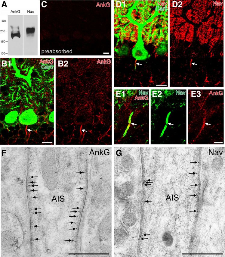
The specificity of AnkG and pan-NaV antibody and their colocalization in the AIS. A, Immunoblot with guinea pig AnkG antibody and rabbit pan-NaV antibody that recognize major bands at 200 or 250 kDa, respectively. B, Double immunofluorescence for AnkG (red; B1, B2), Car8 (green; B1) in the cerebellar cortex. Arrow indicates a PC axon labeled for AnkG. C, Abolished immunofluorescent labeling in the cerebellar cortex with use of preabsorbed AnkG antibody. D, Double immunofluorescence for Nav (red; D1, D2), Car8 (green; D1) in the cerebellar cortex. E, Double immunofluorescence for AnkG (red; E1, E3), Nav (green; E1, E2). Note colabeling in the AIS of PCs (arrow). F, G, Postembedding immunogold electron microscopy showing selective labeling for AnkG (F) and NaV (G) in the membrane undercoating (arrows). Scale bars: B–D, 10 μm; E, 5 μm; F, G, 250 nm.
