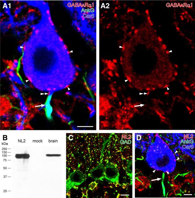
Figure 6.
Immunofluorescence showing the lack of postsynaptic molecules required for GABAergic synapses on the AIS of PCs. A, Triple immunofluorescence for AnkG (green; A1), Car8 (blue; A1), and GABAARα1 (red; A1, A2) in PCs. Note that GABAARα1-immunoreactive clusters are found on Car8-labeled somata or the axon hillock of PCs (arrowheads) but not on the AIS colabeled for AnkG and Car8 (arrows). A doubled arrowhead indicates a GABAARα1 cluster on an AnkG-lacking portion of PC axon. B, Immunoblot showing the specificity of rabbit NL2 antibody. Single protein bands at 100 kDa are detected in HEK cell lysates transfected (left lane) with NL2 cDNA and mouse brain homogenates (right lane) but not in nontransfected HEK cell lysates (mock, middle lane). C, Double immunofluorescence for NL2 (red) and GAD (green). Extensive colocalization in the neuropil and around PC somata supports the specificity of NL2 immunolabeling. D, Triple immunofluorescence for AnkG (green), Car8 (blue), and NL2 (red) in PCs. Note that NL2+ puncta are detected on Car8-labeled PC somata (arrowheads) but not on AnkG-labeled AISs (arrow). Scale bars: 5 μm; C, D, 10 μm.

