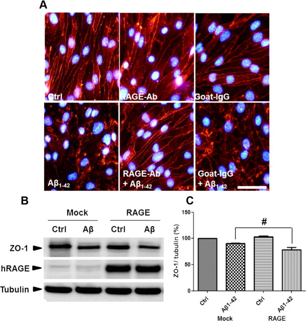
Figure 4.
RAGE mediates Aβ1–42-induced disruptions in TJs. A, Aβ1–42-induced alterations in ZO-1 distribution are attenuated by an anti-RAGE antibody. Con, DMSO; RAGE-Ab, neutralizing anti-RAGE antibody; goat-IgG, IgG from goat used as a negative control. Red signal is ZO-1 staining. Three independent experiments were performed. Scale bar, 100 μm. B, C, Transiently transfected bEnd.3 cells with mock or full-length human RAGE (hRAGE) were incubated with 5 μm Aβ1–42 for 24 h. Representative Western blot images (B) and the densitometry results (C) are presented. Data are represented as mean ± SEM of three independent experiments performed in triplicate. #p < 0.05 versus Mock Aβ1–42-treated sample of RAGE Aβ1–42.

