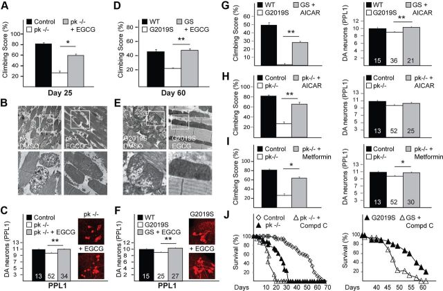
Figure 2.
EGCG treatment mitigates dopaminergic and mitochondrial dysfunction in both mutant LRRK2-expressing and parkin-null flies. A, Climbing score of untreated and EGCG-treated parkin-null (pk−/−) flies, as indicated. B, TEM images of indirect flight muscles of pk−/− flies treated with DMSO or EGCG. C, Quantification of DA neurons in the PPL1 cluster of untreated and EGCG-treated pk−/− flies. Inset, Confocal microscopy images of TH-positive neurons in PPL1 cluster of untreated and EGCG-treated pk−/− flies. D–F, same as A–C, respectively, except that pk−/− flies are substituted with Ddc-driven (D, F) or 24B-driven (E) G2019S LRRK2-expressing flies. Boxed regions in top panels of B and E are shown at higher magnification in corresponding bottom panels. G, Climbing score (left) and DA neuronal count (PPL1 cluster) (right) of Ddc-driven untreated or AICAR-treated LRRK2 G2019S-expressing flies relative to control flies. H, As in G, except that LRRK2 G2019S-expressing flies are substituted with pk−/− flies. I, As in H, except that AICAR is replaced by metformin. Data for control and pk−/− (i.e., climbing score and DA neurons) shown in H and I were derived from A and C. Number indicates the number of fly brains used for DA neuronal quantification. J, Survival curves of untreated or compound C-treated pk−/− (left) or LRRK2 mutant flies (right).

