Figure 1.

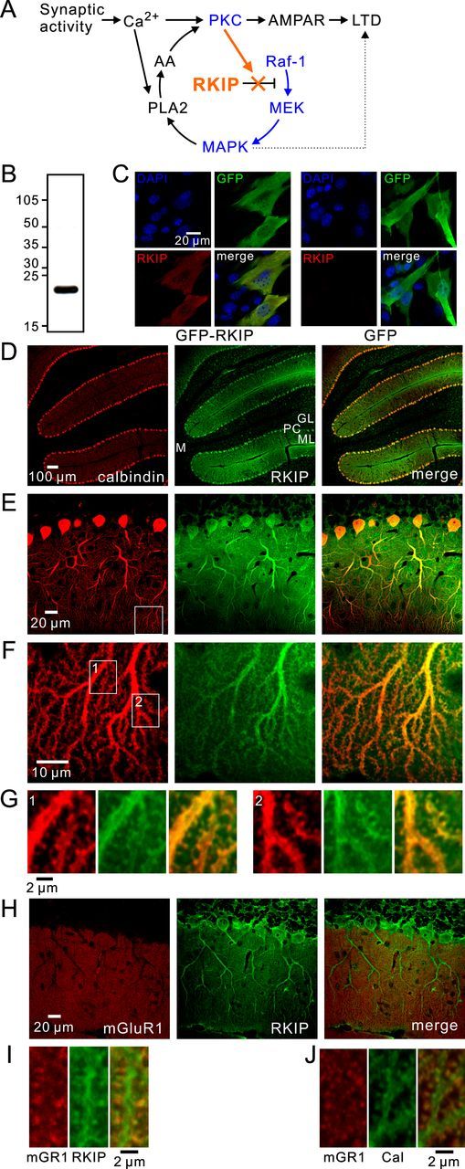
Expression of RKIP in Purkinje cells. A, Proposed model of the involvement of RKIP in the positive feedback loop for LTD (orange). PKC and MAPK pathways are shown in blue. B, Immunoblotting using an antibody against RKIP in cerebellar slices. C, Images of NIH3T3 cells transfected with GFP or GFP-RKIP (green) and stained with a RKIP antibody (red). D–G, Confocal images of a cerebellar slice double stained with antibodies against calbindin (red) and RKIP (green). GL, Granule cell layer; PC, Purkinje cell layer; ML, molecular layer; M, cerebellar medulla. The areas within the white squares shown in E and F are magnified in F and G, respectively. H–J, Confocal images of cerebellar slices double stained with antibodies against mGluR1 (red) and RKIP (green, H, I), or with antibodies against mGluR1 (red) and calbindin (green, J). Magnified images of distal dendrites are shown in I and J.
