Figure 6.

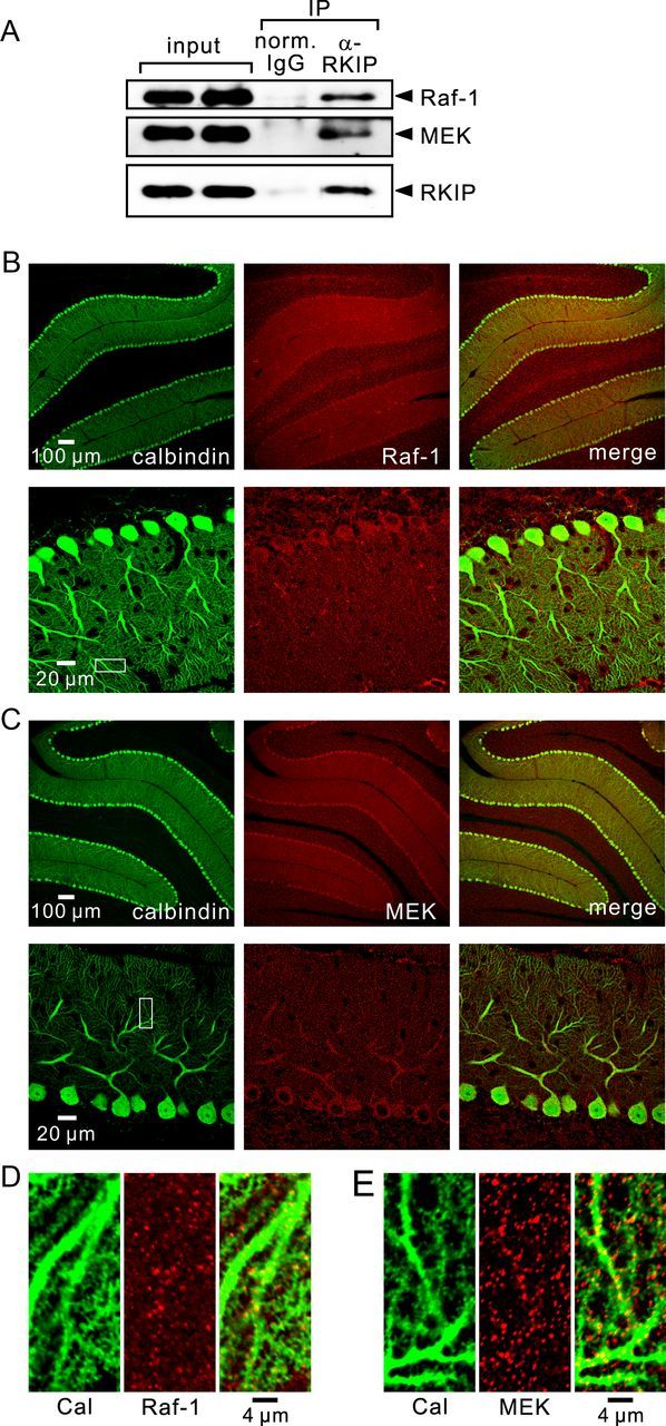
Interaction of Raf-1 and MEK with RKIP, and localization of Raf-1 and MEK in cerebellar slices. A, Immunoblot analysis of coimmunoprecipitated Raf-1, coimmunoprecipitated MEK, and precipitated RKIP in lysates from control cerebellar slices. B–E, Confocal images of cerebellar slices double stained with antibodies against calbindin (green) and Raf-1 (red; B, D), or with antibodies against calbindin (green) and MEK (red; C, E). Areas within the white squares shown in B and C are magnified in D and E, respectively.
