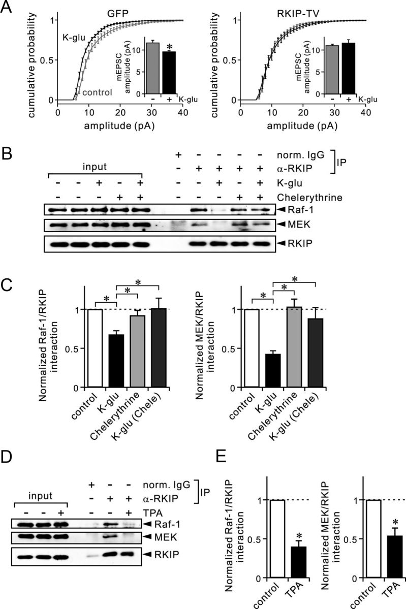
Figure 7.

Dissociation of RKIP from Raf-1 and MEK during chemical LTD stimulation depends on PKC. A, Summary of mEPSC amplitudes in Purkinje cells, shown as cumulative distributions and bar graphs (inset). Results were obtained by recording from 10 (control) or 8 (K-glu) cells expressing GFP (left), and by recording from 12 (control) or 8 (K-glu) cells expressing GFP-RKIP-TV (right). B, Immunoblot analysis of coimmunoprecipitated Raf-1, coimmunoprecipitated MEK, and precipitated RKIP in cerebellar slices, which were treated with or without K-glu in the absence or presence of chelerythrine (5 μm). C, Quantification of the interaction between RKIP and Raf-1 (left) or between RKIP and MEK (right) in cerebellar slices. Control slices were incubated only in normal ACSF. Other slices were treated with K-glu or chelerythrine as indicated. Results were obtained from 21 (K-glu only) or 11 (chelerythrine) experiments for Raf-1, and from 4 experiments for MEK. D, Immunoblot analysis of coimmunoprecipitated Raf-1, coimmunoprecipitated MEK, and precipitated RKIP in cerebellar slices, which were treated with or without TPA (0.5 μm). E, Quantification of the interaction of RKIP with Raf-1 (left) or MEK (right) in cerebellar slices, which were treated with or without TPA. Results were obtained from five experiments. *p < 0.05 (A); *p < 0.01 (C, E) [Student's t test (A, E); ANOVA followed by the Fisher's PLSD test (C)]. Ratios of coimmunoprecipitated Raf-1 or MEK to precipitated RKIP shown in C and E were normalized with the ratio of the control condition. Error bars represent SEM.
