Figure 2.
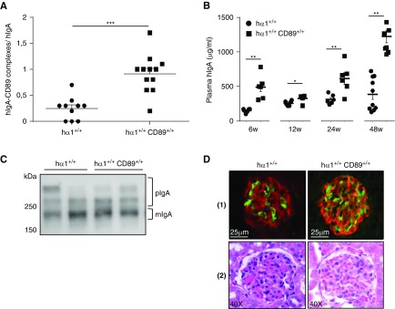
Coexpression of human CD89 and IgA does not affect IgA toxicity in mice. (A) Plasma hIgA-CD89 levels in hα1+/+ and hα1+/+CD89+/+ mice at 12 weeks. OD of (hIgA-CD89 complexes) was normalized to OD of hIgA in each model (n=10–12 mice). (B) Plasma hIgA levels in mice at 6, 12, 24, and 48 weeks. (C) Analysis of hIgA circulating forms by western blot at 48 weeks. Blots are representative for n=6 mice per condition; two mice from each group are presented. (D, 1) hIgA deposits are in green. (D, 2) Hematoxylin and eosin staining of kidney sections from mice at 24 weeks. Pictures are representative for all glomeruli found in kidney sections of n=6–12 mice per condition. For numeric data, results are means ± SEM of n=6–10 mice per group. hIgA, human IgA. *P<0.05; **P<0.01; ***P<0.001.

