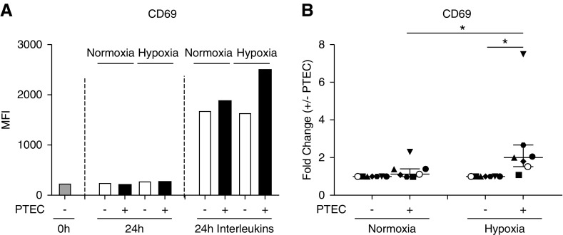
Figure 6.
MAIT cells activated in the presence of hypoxic PTECs display significantly upregulated expression of CD69. (A) CD69 expression by MAIT cells freshly isolated (0 hours) or after 24-hour culture without (−) or with (+) preconditioned PTECs under normoxic or hypoxic conditions in the absence (24-hour) or presence (24-hour IL) of IL-12p70, IL-15, and IL-18. Surface expression was measured by flow cytometry (gated on live, single, CD45+ cells) and expressed as the median fluorescence intensity (MFI). One representative donor experiment is shown. (B) Fold changes (MFI in the presence of PTECs/MFI in the absence of PTECs; +/− PTECs) in CD69 levels on IL-stimulated MAIT cells under normoxic and hypoxic conditions for seven individual donor PTEC experiments. Symbols represent individual donor PTEC experiments; the representative donor experiment from (A) is identified using open circles. Horizontal bars represent medians, with interquartile range also presented. *P<0.05, Wilcoxon matched-pairs signed-rank test.

