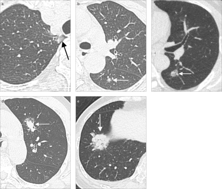
Figure 2. a–e.
Part-solid nodules of various sizes that correspond to different T stages. Chest CT images (a, b) of a 55-year-old female with two part-solid nodules, one found in the apical segment of the right upper lobe of the lung with total diameter of 1.0 cm and a solid component of 0.3 cm (a, arrow), and the other noted in the superior segment of the left lower lobe of the lung with a diameter of 0.7 cm and a solid component of 0.4 cm (b, arrow). Pathology proved adenocarcinoma of lung. The imaging staging was cT1mi (m), m for multiple nodules. CT image (c) of a part-solid nodule with a solid component measuring up to 0.8 cm in maximum diameter with total size of 1.8 cm (arrow), imaging staging T1a, in a 53-year-old man with pathology showing non-small cell carcinoma. Lung CT image (d) of a 62-year-old male, a case of esophageal cancer, pT2N0M0, status post thoracoscopic esophagectomy and gastric tube reconstruction. Follow-up chest CT found one part-solid nodule up to 1.8 cm with 1.2 cm solid part (arrow) in the left upper lobe without obvious lymph node and distant metastasis, imaging staging T1a (≤2 cm) in the 7th edition and T1b in the 8th edition, suspected primary lung cancer. Pathology showed moderately differentiated mixed mucinous and acinar adenocarcinoma and hilar/mediastinum lymph node metastasis. Lung CT image (e) of a 76-year-old female with incidental finding of a 2.8 cm part-solid tumor with a solid part measuring up to 2.7 cm (arrow), located at the right lower lobe of the lung. Imaging staging of AJCC 8th edition is T1c. CT-guided biopsy revealed adenocarcinoma.

