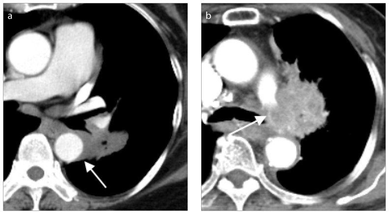
Figure 5. a, b.
Lung cancer with major vessel invasions. Lung cancer at superior segment of the left lower lobe (a, arrow), with maximum tumor diameter of 4 cm, in a 71-year-old female with pathology from endobronchial ultrasound biopsy proving non-small cell carcinoma, favoring adenocarcinoma. Lung CT showed tumor invasion of descending aorta with contact length more than one fourth (90°) of the circumference of descending aorta, and thus increased the T classification to T4, according to the 8th edition of lung cancer staging. Chest CT (b) showing lung cancer (arrow), with maximum diameter of 4.9 cm, in an 83-year-old female with tumor invasion to the left pulmonary artery.

