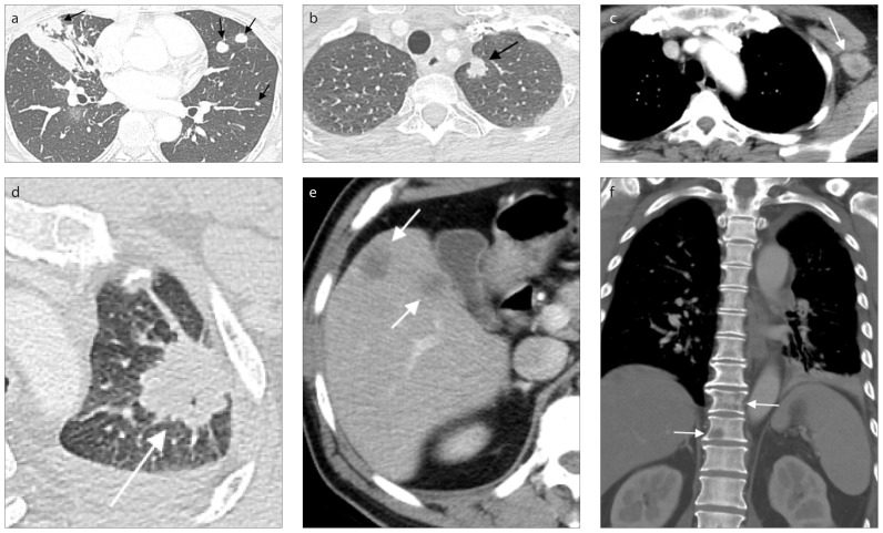
Figure 6. a–f.
CT scans showing three classifications of M descriptors. Chest CT image (a) of a 51-year-old female with a mass having maximum diameter of 4.2 cm and multiple pulmonary nodules in bilateral lungs (arrows), suspected bilateral lung to lung metastasis. Biopsy pathology favored primary lung adenocarcinoma. This case was staged M1a in image staging, with intrapulmonary metastasis. Contrast-enhanced chest CT images (b, c) of a 54 year-old female with lung tumor at left upper lobe of lung and distal metastasis to a nonregional lymph node. Lung window (b) showing the 1.3 cm solid tumor nodule with spiculated margin in the apicoposterior segment of left upper lobe of lung (arrow). Soft tissue window (c) illustrating the extrathoracic lymph node metastasis at left axillary region (arrow). Since no other distant metastasis is detected, this case should be staged as M1b, a single extrathoracic metastasis in a single organ. A case of M1c classification (d–f): multiple extrathoracic metastatic lesions at multiple sites of a 61-year-old male with lung cancer in the apicoposterior segment of left upper lobe of lung with a diameter of 3.3 cm (d, arrow). Soft tissue window (e) showing poorly enhanced nodules (arrows) in segment VII, VIII and V of liver, suspected metastatic lesions. Bone metastatic osteolytic lesions were noted in T10 and T11 vertebrae (f, arrows).

