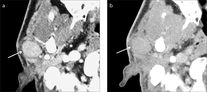
Figure 4. a, b.
Warthin tumor of the right parotid gland in a 50-year-old man. A well-defined ovoid mass (arrow) is seen in the superficial lobe of the right parotid gland on two-phase CT scans. The mass shows marked homogeneous contrast enhancement at early phase (a) and homogeneous washout of contrast material at delayed phase (b). The measured AE and AD are 121 and 90, respectively, resulting in AWO of 31 and RPEWR of 25.6%.

