
Figure 3. DNA vaccine–primed T cells selectively kill mutated cells.
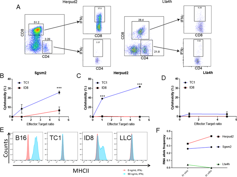
A. Representative flow cytometry plots showing IFNγ-expressing CD4+ and CD8+ T cells resulting from the expansion of T cells from mice immunized with TC1 plasmid 1 or TC1 plasmid 2, stimulated with Herpud2 or Lta4h peptides. Negative control: non-peptide stimulated T cells (n=5 mice/group). B-D. Cytotoxicity of T cells expanded from mice immunized with TC1 plasmids (from A). T cells were expanded with Sgsm2 (B), Herpud2 (C) or Lta4h (D) specific peptides (5μg/mL). After expansion, T cells were cocultured with 10,000 luciferase-tagged TC1 or ID8 tumor cells. Cytotoxicity was measured by luciferase activity after 24 hours of coculture. E. RNA expression Sgsm2, Herpud2 and Lta4h in TC1 tumor cells grown in vivo and in vitro (2 mice/tumor type after 3 weeks). F. Representative flow cytometry histograms showing surface expression of MHC class II on B16, TC1, ID8, and LLC tumor cells in normal conditions or after being exposed to IFNγ (50ng/mL) for 48 hours. (Single experiment). Two-way ANOVA. ***p<0.001.