
Figure 2. Caspase-3-mediated unconventional secretion of NS1 during MNoV infection.
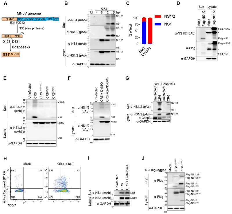
(A) Schematic depicting NS1 protein maturation. (B) Immunoblots showing NS1 secretion during CR6 infection in BV2 cells. (C) Relative expression of NS1 and NS1/2 in the supernatant (sup) and in the lysate. Band intensity of immunoblots at 12 hpi was measured from five independent experiments. Shown are means ± SD. (D) HEK293T cells were transfected with Flag-NS1/2-CR6, and Flag-NS1 secretion was examined 48 hours after transfection. (E) NS1 secretion at 12 hpi with Caspase-cleavage site mutant viruses. (F) BV2 cells were treated with Q-VD-OPh (20 nM) at 4 hpi and NS1 secretion was examined at 12 hpi. (G) Casp3KO BV2 cells were generated by CRISPR, and were infected with CR6 to access NS1 secretion. (H) Intracellular flow cytometry plots. CR6-infected BV2 cells were co-stained with cleaved-Casp3 and NS6/7 at 16 hpi. (I) Brefeldin A (5 ng/ml) treatment to block the conventional secretion pathway. The cells were incubated with Brefeldin A from 4 hpi and NS1 secretion was analyzed at 12 hpi. (J) HEK293T cells were transfected with Flag-NS1/2CR6 or Flag-NS1/2GI.I and Flag-NS1GI.1 cleavage and secretion was detected in the sup. Asterisk means non-specific bands. Representative data from at least three experiments are shown, except (I). See also Figure S2.