
Figure 5. The defective viral growth of CR6D121/131G is complemented in IFN-λ signaling deficient mice.
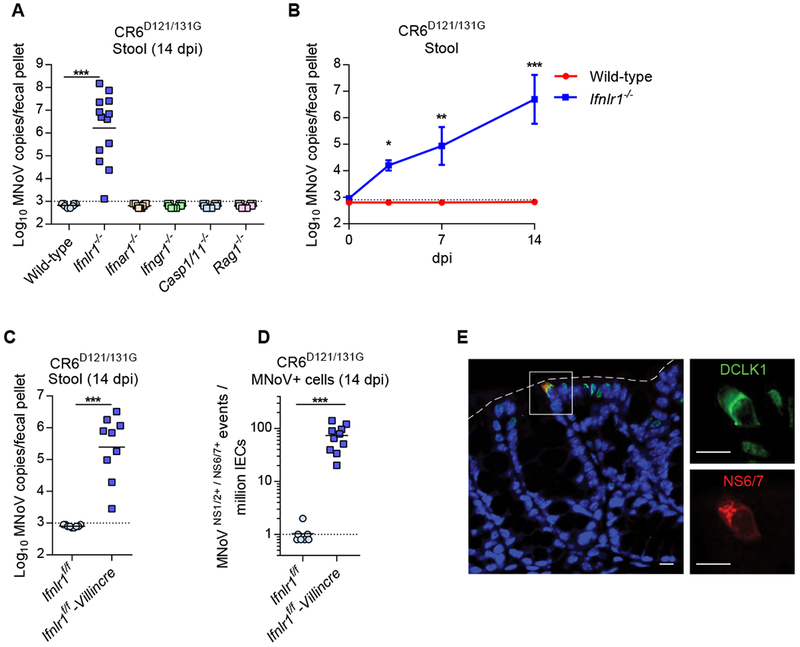
(A-E) Wild-type and the knock out mice were infected with 107 PFU of CR6D121/131G perorally. (A) Complemented viral infection of CR6D121/131G in Ifnlr1−/− mice (n = 9-13, combined from three independent experiments). (B) Viral shedding of CR6D121/131G at 3, 7 and 14 dpi in Ifnlr1−/− mice (n = 5-6, combined from two independent experiments). (C) CR6D121/131G stool shedding in Ifnlr1f/f-Villincre mice. (D) Quantification of MNoV+ cells (NS1/2+NS6/7+) in IECs (CD45−EpCAM+) by flow cytometry. (E-F) n = 9-10, combined from two independent experiments. (E) MNoV-NS6/7 co-localizes with DCLK1, a tuft cell marker, in the colon of Ifnlr1−/− mice infected with CR6D121/131G at 14 dpi. Images are representative of one of at least three independent experiments. Dashed lines represent the epithelial barrier. White boxes in the overlaid image reflect the magnified inset images. Scale bars, 10 microns. *P < 0.05; **P < 0.01; ***P < 0.001, determined by Mann-Whitney test (A, C, and D) or two-way ANOVA (B).