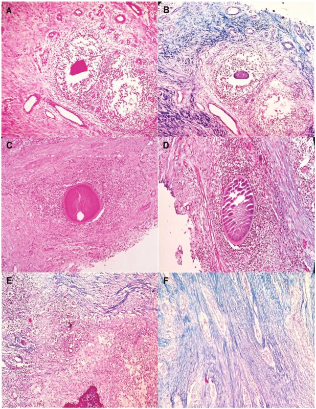
Fig 2. Collagen deposition around different mycetoma causatives agent; in this figure a representative picture of the A. pelletieri grain insight the subcutaneous tissue from one particular patient is shown.
In panel A, a HE staining is performed. In panel B, a Masson trichrome staining of the same area is shown. As is seen on this slide, collagen (coloured blue) is mainly seen within zone 2 as a fine thin bundles that surrounded the grains. In panel C, a HE staining is performed from patient with S. somaliensis; and D, collagen fibers is mainly seen within zone 2 as a fine thin bundles that surrounded the grains; in Panels E and F showed a collagen fibers in M. mycetomatis, with thick collagen bundles. This figure is composed of representative pictures from single patients. Differences among individual patients were noted, however we chose pictures which correlated with the majority of each group of patients.

