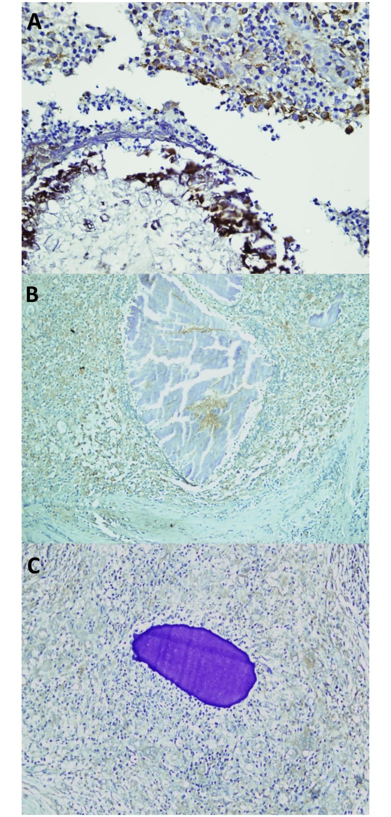
Fig 3. IL-17A expression in M. mycetomatis (A), S. somaliensis (B) and A. pelletieri (C) lesions.

IL-17A staining was seen as a brown cytoplasmic staining in cells. This figure is composed of representative pictures from single patients. Differences among individual patients were noted, however we chose pictures which correlated with the majority of each group of patients.
