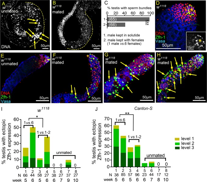
Fig 1. Reproduction induces ectopic Zfh-1 expression away from the hub.
(A and B) Basal ends of 5w-old w1118 testes stained for DNA to visualize the bundles of spermatid nuclei (Hoescht 33342). (A) Testis from males kept in solitude. Sperm bundles are indicated by arrows. (B) Testis from a mated male (1 male vs. 6 females). There was no sperm bundle at the basal end. (C) Percentages of the testes containing at least one sperm bundle. (D-H) Testes from w1118 males immunostained for Zfh-1 (CySC/early cyst cell, green), co-stained for Vasa (germ cells, blue) and DNA (nuclei, red). Asterisks indicate testis hubs. (D) Apical region of a 1-3-day-old testis. Zfh-1-positive cells and small, DNA-bright Vasa-positive early germ cells were only present in the apical region. Inset shows the Zfh-1 expression in the tip. (E-H) 5w-old testes from the males kept in solitude (unmated) (E), or from single males mated with six females (1 vs. 6) (F-H). Ectopic Zfh-1-positive cells are indicated by arrows in the testes exhibiting level 1 (F), level 2 (G) and level 3 (H) ectopic Zfh-1 expression. In level 2 (G) and level 3 (H) testes, ectopic Zfh-1-positive cells were usually associated with small germ cells. (I and J) Percentages of w1118 (I) and Canton-S (J) testes with ectopic Zfh-1 expression. Extra Zfh-1-positive cells (levels 1–3) were observed in testes from single male mated with 6 females (1 vs. 6) and 1–2 males (1 vs. 1–2) but rarely seen in the testes from unmated males. The asterisks indicate the significance comparing total testes from week-5 and -6 males in 1 vs. 6 to 1 vs. 1–2 mating ratio. N = number of testes scored. P-values shown in C, I, and J were calculated with Chi-squared test. *p<0.05, **p<0.01, and ***p<0.001.

