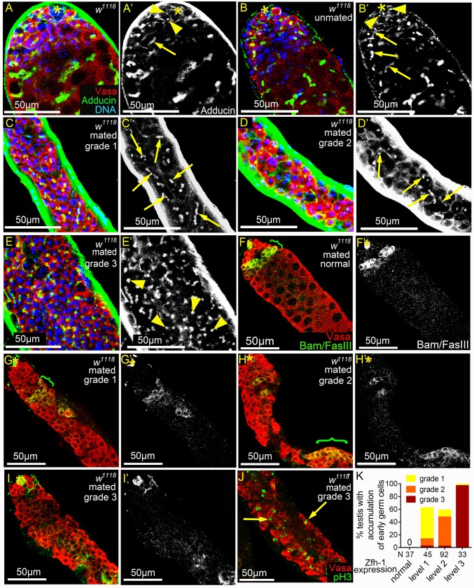
Fig 3. Reproduction leads to early germ cell accumulation and reduced Bam expression.
(A-E’) w1118 testes immunostained for Adducin (green, white in A’-E’), co-stained for Vasa (red) and DNA (blue). Dot spectrosomes and thin fusomes are indicated by arrowheads and arrows, respectively. Asterisks mark the hubs. (A-B’) Dot spectrosomes and thin fusomes were observed only in the apical region of the testes from 1-3-day-old (A and A’) and 5w-old unmated (B and B’) males. (C-D’) Thin fusomes were found in the middle part of the grade 1 (arrows in C and C’) and grade 2 (arrows D and D’) testes that exhibit excess early germ cells. (E and E’) Dot-like spectrosomes (arrowheads) were observed away from the hub in the grade 3 testes. (F-I’) Testes from mated 5w-old w1118 males immunostained for Bam and FasIII (green in F-I and white in F’-I’), co-stained for Vasa (red). Bam-positive regions are indicated with brackets. Asterisks mark the hubs. (G-I’) Testes with expansion of early germ cells. Bam expression was markedly reduced and the onset of Bam expression was delayed in grade 1 and 2 testes (G and H). Anti-Bam signals were not detected in grade 3 testes (I). (J) Testis from mated 5w-old w1118 male immunostained for pH3 (green), co-stained for Vasa (red). Arrows indicate the single germ cells undergoing mitosis. (K) Percentages of mated 5w-old w1118 testes exhibiting excess small germ cells. The percentages and severity of accumulation of early germ cells (Y-axis) were positively correlated with the degrees of ectopic Zfh-1 expression (X-axis). Mass-mating of 10 males and 20 females was conducted for all experiments.

