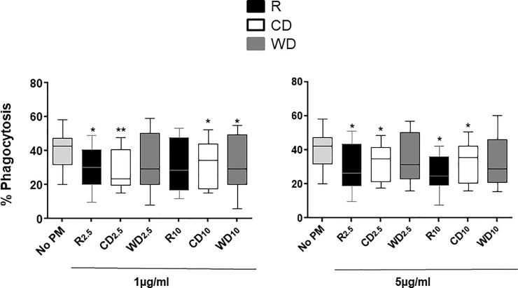
Fig 2. PM effects on M.tb phagocytosis.
MDM were exposed to 1 and 5 μg/ml of R2.5 (n = 10, 1 μg/ml; n = 8, 5 μg/ml), CD2.5 (n = 9), WD2.5 (n = 9), R10 (n = 9, 1 μg/ml; n = 7, 5 μg/ml), CD10 (n = 9), and WD10 (n = 9) or left unexposed (n = 11) at 37ºC, 5% CO2 in a humidified environment for 18 h. Following exposure to PM, MDM were infected with M.tb at MOI1 for 2 h. Phagocytosis of M.tb by MDM was assessed by identification of acid-fast bacilli (Materials and methods). Proportions of MDM with intracellular M.tb were determined by bright field microscopy (1000x, oil immersion) within a total of 300 MDM in each experimental condition. Data are presented as medians, interquartile ranges (IQR) and 5th and 95th percentiles. Statistical comparisons were done by non-parametric Wilcoxon matched pair signed rank test. Statistically significant changes relative to M.tb-infected (no PM control) PBMC within each dose are shown with * (p ≤0.05) or ** (p ≤0.001).

