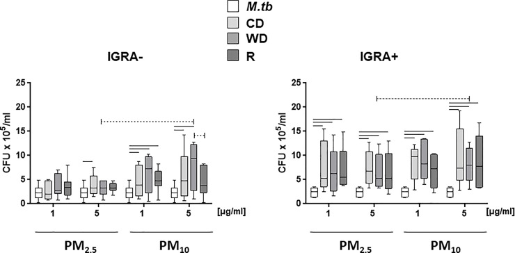
Fig 6. Effect of PM size and seasonal source on M.tb growth control.
PBMC from IGRA- (n = 7) or IGRA+ (n = 5) subjects were exposed to 0 (no PM), 1 and 5 μg/ml of R2.5, CD2.5, WD2.5, R10, CD10, or WD10 at 37ºC, 5% CO2 in a humidified environment for 20 h. Following pre-exposure to PM, PBMC were infected with M.tb at MOI1 or left uninfected. After 2 h infection, non-phagocytosed M.tb was removed by washing and plates subsequently incubated in complete culture media. PBMC were lysed and serial dilutions of cell lysates plated in M.tb growth media in triplicate on 7H10 agar plates and incubated at 37°C for 21 days until M.tb colony forming units (cfu) were determined. Results from day 4 are shown. Statistically significant (p≤0.05) differences relative to PM-unexposed M.tb-infected control PBMC are shown with solid lines. Size and season-specific differences are shown with dotted lines.

