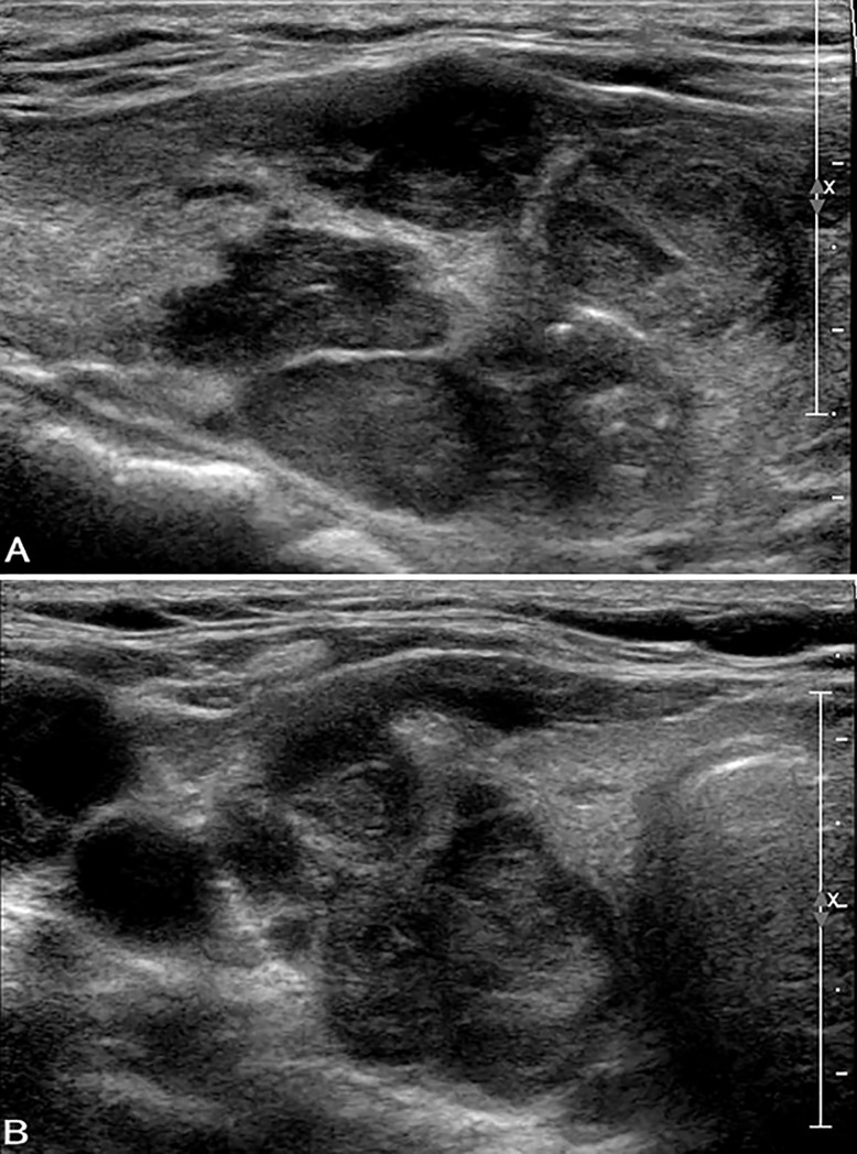
Fig 3. Ultrasound images in a patient with FTC (widely invasive) in the right lobe.
A, B. Longitudinal (A) and transverse (B) US show a hypoechoic solid thyroid nodule (size: 43 mm × 24 mm × 29 mm) with wider-than-tall shape and extra-thyroidal extension. Macrocalcification and punctuate echogenic foci are observed. The nodule was classified as ACR TIRADS category TR5. The cytological diagnosis was follicular neoplasm with positive K-RAS mutation.

