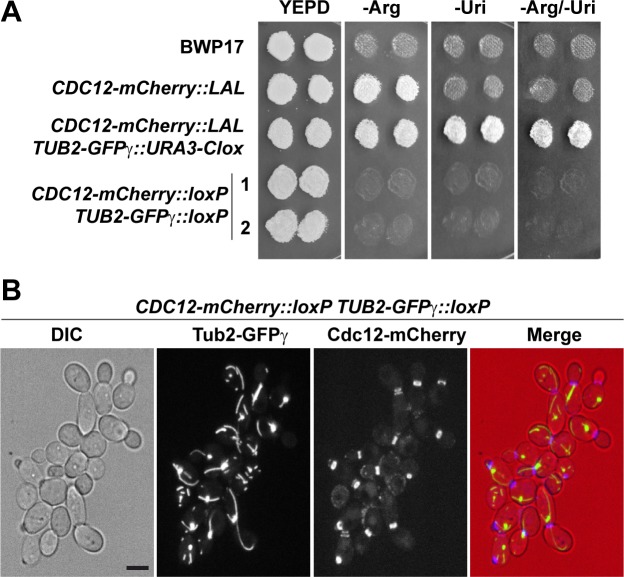
Fig 4. Double fluorescent tagging with GFPγ and mCherry.
(A) Growth in selective media. Single colonies of strains BWP-17, CDC12-mCherry::LAL, CDC12-mCherry::LAL TUB2-GFPγ::URA3-Clox and two independent clones of the resolved strain CDC12-mCherry::loxP TUB2-GFPγ::loxP (1 and 2) were grown on YPD plates and replica-printed to SC -Arg, SC -Uri or SC -Arg/-Uri media. (B) Localization of Cdc12-mCherry and Tub2-GFPγ during yeast growth. Exponential growing cultures of the CDC12-mCherry::loxP TUB2-GFPγ::loxP strain were imaged. The images are the maximum projection of 5 planes and show the differential interference contrast image (DIC), the Tub2-GFPγ and the Cdc12-mCherry channels, and the merged image (DIC in red, Tub2-GFPγ in green and Cdc12-mCherry in blue). Scale bar, 2 μm.

