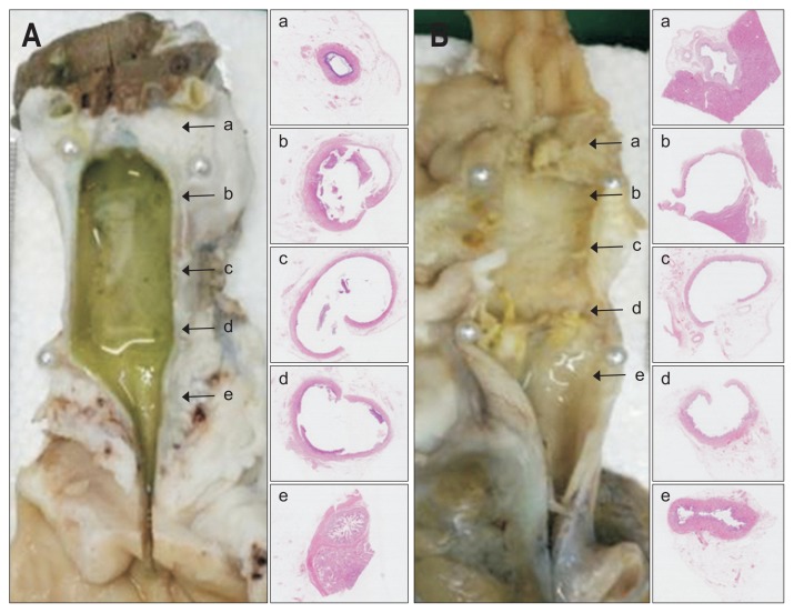
Fig. 2.
Histology of extracted common bile ducts showing similar inflammatory and fibrotic reactions after stenting with 0% paclitaxel (A) or 10% paclitaxel (B). Gross appearance (left panel) and corresponding microscopic views (right panel: H&E, ×12.5), arrows indicate areas sampled for slide preparation. Proximal (a) and distal (e) portions of non-stented areas retain villous mucosal pattern in both groups, whereas proximal (b), central (c), and distal (d) portions of stented areas display luminal dilatation, mucosal atrophy, and attenuated walls.

