Figure 10.

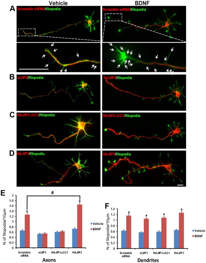
JIP3 regulates axonal filopodia formation in response to BDNF in hippocampal neurons. After staining with Alexa 488-phalloidin, filopodia were defined as any protrusion under 10 μm in length. The number of filopodia and the length of axonal or dendritic shaft were then computed to obtain the filopodia density (number of filopodia per 10 μm). A–D, Hippocampal neurons (5 DIV) transfected with scramble siRNA (A), siJIP3 (B), HAJIP3ΔCC1(C), or HAJIP3 (D) were treated with BDNF (100 ng/ml) for 20 min, with the unstimulated group as vehicle control. Existence of transfected siRNA could be detected by RFP fluorescence; the expression of HAJIP3 or HAJIP3ΔCC1 was detected by immunoblotting with HA antibodies (red). Bottom panels in A showed enlarged images of the framed regions with arrows highlighting the filopodia. E, F, The number of filopodia in axons (E) or dendrites (F) per 10 μm were calculated. For each neuron, the length of axon or dendrite length ranging between 30 and 80 μm was analyzed. The results of more than three independent experiments were compiled. Results are presented as the mean ± SEM (*p < 0.05, vs the vehicle control; #p < 0.05, vs scramble siRNA group stimulated with BDNF; one-way ANOVA). Scale bars: A, D, 10 μm.
