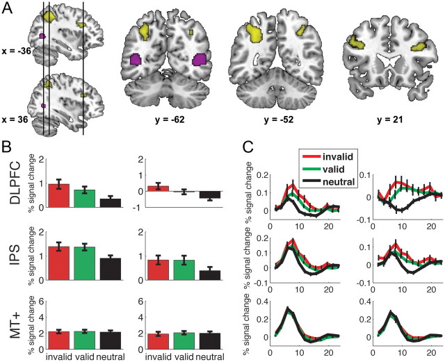
Figure 3.
Neural activation differences induced by expectation. A, Larger activation was found bilaterally in both IPS and bilateral DLPFC for trials in which subjects had an expectation than for trials in which subjects had no expectation (shown in yellow). Bilateral MT+ (shown in purple) was functionally localized using an independent localizer for each subject. B, Percentage signal change is plotted for each of the three cue types (invalid, valid, and neutral) for left DLPFC, IPS, and MT+ (left column) and its right hemisphere counterpart (right column). DLPFC showed larger activity for invalidly cued trials compared with validly cued trials (both p values <0.02). No such difference was found for IPS (both p values >0.8). There were no differences in MT+ for the differently cued trials (all p values >0.2). C, Time courses for each of the six regions of interest are plotted for each trial type. Error bars represent the SEM.

