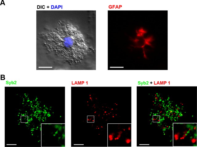
Figure 2.
Small vesicles and lysosomes in freshly isolated astrocytes. A, Representative images showing a freshly isolated astrocyte. Left, DIC image and DAPI (blue) staining. Right, GFAP staining (n = 17 cells). Scale bars, 10 μm. B, Double immunostaining of Syb2 (green; left) and LAMP1 (red; middle) in another freshly isolated astrocyte. Merged image at right. Syb2 did not colocalize with LAMP1 in freshly isolated astrocytes. Scale bars, 10 μm. The boxed areas are magnified in the insets.

