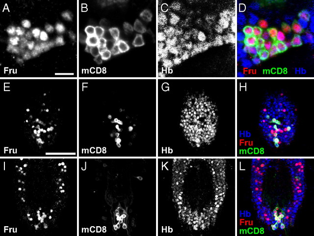
Figure 2.
Coexpression of Hb and Fru in the postembryonic CNS. A–D, Images of mAL neurons in the pupal male brain stained with the anti-Fru antibody (A; red in D), the anti-mCD8 antibody for fruNP21 expression (B; green in D) and the anti-Hb antibody (C; blue in D); and the merged image (D). Scale bar, 10 μm. E–L, The ventral (E–H) and dorsal (I–L) parts of the ventral nerve cord at 24 APF stained with the anti-Fru antibody (E, I; red in H and L), and the anti-mCD8 antibody for fruNP21 expression (F, J; green in H and L) and the anti-Hb antibody (G, K; blue in H and L); the merged images are shown in H and L. Scale bar, 50 μm.

