
Figure 4.
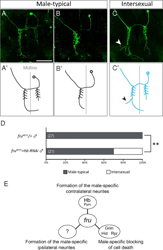
Demasculinization of the contralateral neurite structure of mAL detected in single-cell MARCM clones. A–C′, Three different examples of single-cell clones of mAL are shown. The original images (A–C) and schematic drawings (A′, B′, C′) of mAL neurons are shown. The female-type side branch is highlighted with an arrowhead. Scale bar, 50 μm. D, The proportion of flies that carried single-cell clones of male-type mAL (p < 0.01 by Fisher's exact test). The genotype of the test flies was: y hs-flp/Y;G13 UAS-mCD8-GFP/G13 tub-Gal80;fruNP21 UAS-dicer2/UAS-hb RNAi. E, Three distinct actions of Fru in producing sexual dimorphism in three different characteristics of mAL neurons.