
Figure 3.
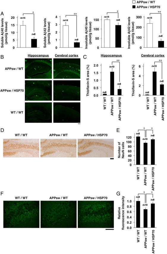
Effects of HSP70 overexpression on Aβ levels, Aβ plaque deposition, and neuronal and synaptic loss in the brain of transgenic mice expressing APPsw. A, Soluble and insoluble fractions were prepared from the brains of 12-month-old APPsw/WT (n = 14) and APPsw/HSP70 mice (n = 8). The amounts of Aβ40 and Aβ42 in each fraction were determined by sELISA as described in Materials and Methods. B, D, F, Brain sections were prepared from 18-month-old APPsw/WT (n = 10), APPsw/HSP70 (n = 8), and WT/WT mice (n = 8), and then subjected to thioflavin-S staining (B) and immunohistochemical analysis with an antibody to NeuN (D) or synaptophysin (F). Scale bars: B, 200 μm; D, 100 μm; F, 50 μm. C, E, G, Relative area stained with thioflavin-S (C), number of NeuN-positive cells in hippocampal CA3 region (E), and relative fluorescence intensity (synaptophysin) in hippocampal CA3 region (G) (3 sections per brain) were determined. Values are given as mean ± SEM. **p < 0.01; *p < 0.05.