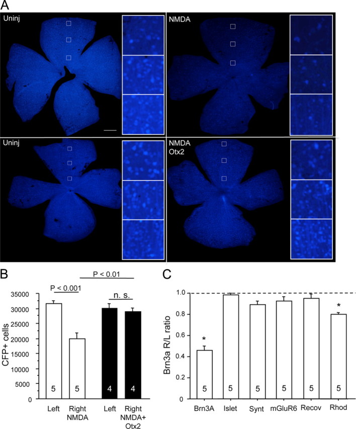
Figure 3.

NMDA excitotoxicity of RGCs. A, Four days after intraocular injection of NMDA (2 mm final) in B6.Cg(Thy1-CFP)23Jrs/j mice, the number of CFP-positive RGC profiles appeared reduced throughout the retina compared to the uninjected (Uninj) retina (top). When Otx2 (30 ng) was injected at the time of NMDA, fewer CFP+ cells appeared lost (bottom). Scale bar, 500 μm. B, Counting CFP-positive RGCs in the experiment depicted in A showed a significant reduction of about 33% compared to the uninjected left eye (2-tailed t test). Thirty nanograms of Otx2 fully protected fully against the NMDA-induced loss of CFP+ RGCs. C, NMDA (2 mm final) significantly reduced Brn3a mRNA, while mRNAs specific for other cell types were little or not affected. (*p < 0.005, one sample t test against null hypothesis that the right eye to left eye ratio = 1.0). The numbers at the bottom of each bar represent the number of mice in each condition.
