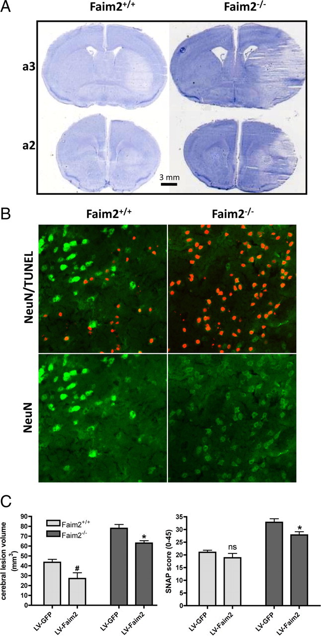
Figure 4.

Effects of transient brain ischemia in Faim2−/− and littermate Faim2+/+ mice. Analyses were performed after 30 min of MCAo and 72 h of reperfusion. n = 8–11 male mice per group, age-matched. Data are expressed as mean ± SEM. Unpaired t test (Prism Software, SigmaStat). A, Examples of proximate (a2, a3) hematoxylin-stained 20 μm coronal brain sections taken from anterior–posterior serial coronal cryostat sections (20 μm). B, Examples of NeuN and NeuN/TUNEL-immunofluorescence staining in infarcted area. C, Stereotaxic injections of LV-Faim2 into the striatum 3 weeks before MCAo reduced lesion size and clinical deficits. SNAP score was evaluated before and 2 h after MCAo. Two-way ANOVA followed by unpaired t test (Prism Software, SigmaStat) was used. Data are shown as mean ± SEM. N = 5–8 animals per group. Lesion volume, Genotype p < 0.0001, virus treatment p = 0.0013, genotype × virus treatment interaction p = 0.8686. #p = 0.0218 versus Faim2+/+ littermates with lentiviral GFP infection, *p = 0.0126 versus Faim2−/− mice with lentiviral GFP infection. SNAP, Genotype p < 0.0001, virus treatment p = 0.0135, genotype × virus treatment interaction p = 0.2955. n.s. (p = 0.54) versus Faim2+/+ littermates with lentiviral GFP infection, *p = 0.0258 versus Faim2−/− mice with lentiviral GFP infection.
