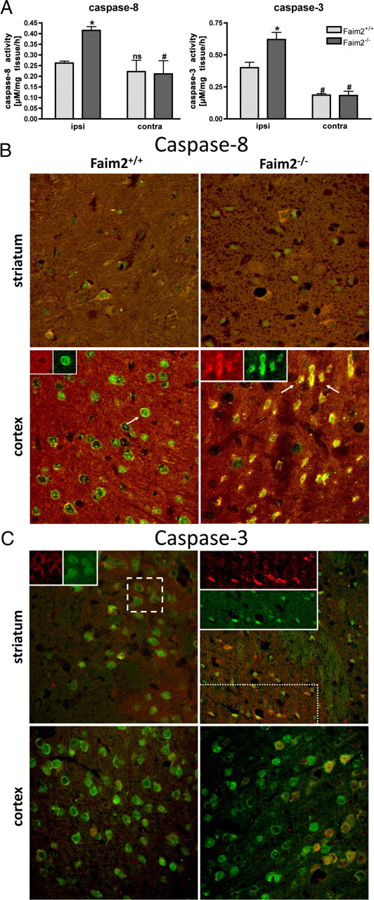
Figure 5.

Caspase-8 and caspase-3 activity in Faim2−/− and littermate Faim2+/+ mice after focal brain ischemia. A, Caspase activities ex vivo in Faim2+/+ and Faim2−/− mice 20 h after 30 min MCAO. Data are shown as means ± SEM pooled from n = 3 animals per condition. Caspase-8, Two-way ANOVA followed by Tukey post hoc analysis (Prism Software, SigmaStat): genotype, F(1,11) p = 0.024; side, p < 0.001; genotype × side interaction, p = 0.021 (caspase-8). *p = 0.004 versus ipsilateral Faim2+/+. #p = 0.01 versus corresponding ipsilateral in Faim2−/− and Faim2+/+ mice. Caspase-3, Two-way ANOVA followed by Tukey post hoc analysis (Prism Software, SigmaStat): genotype, F(1,11) p = 0.129; side, F(1,11) p = 0.020; genotype × side interaction, p = 0.087 (caspase-3). *p = 0.033 versus ipsilateral Faim2+/+. #p = 0.009 versus ipsilateral Faim2−/−. n.s., Not significant versus ipsilateral Faim2+/+. B, Confocal immunofluorescence 16 h after 30 min MCAo. Costaining of NeuN (green) and cleaved caspase-8/subunit 18 (red) (picture size: 200 × 200 μm). C, Confocal immunofluorescence 16 h after 30 min MCAo. Costaining of NeuN (green) and cleaved caspase-3 (red) (picture size: 275 × 275 μm).
