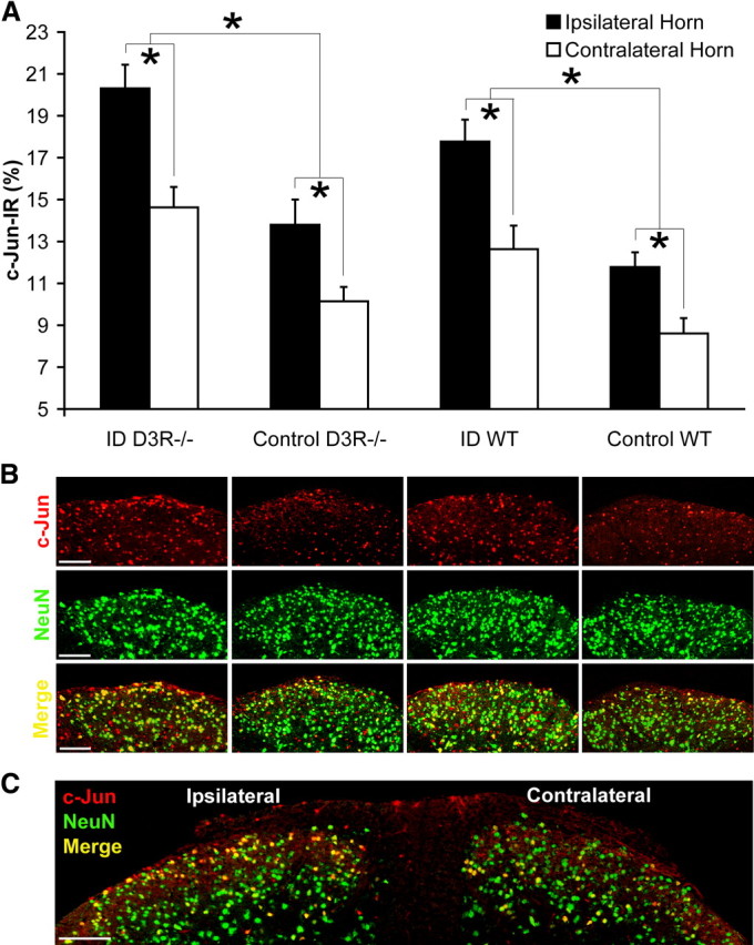
Figure 4.

Altered c-Jun expression at the ipsilateral dorsal horn after formalin-induced pain. A, Percentages of c-Jun-IR neurons were quantified in relation to total neuron count in laminae I/II at either the ipsilateral or contralateral dorsal horns from WT and D3R−/− mouse strains fed control or ID diets for 15 weeks starting from age P28. Values are means ± SEM, n = 7. *WT and D3R−/− groups differ in the percentage level of c-Jun-IR expression, p < 0.05. B, Colocalization (yellow) of c-Jun immunoreactivity (red) in relation to the neuronal marker NeuN (green), at laminae I/II of the ipsilateral dorsal horn (scale bar, 75 μm). C, c-Jun expression is increased at the ipsilateral dorsal horn of an ID D3R−/− mouse (scale bar, 100 μm).
