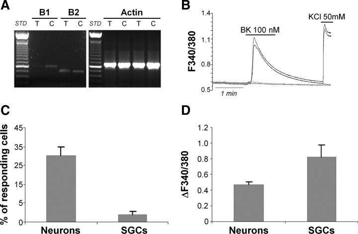
Figure 2.
Trigeminal neurons in vitro express functional BK receptors. A, RT-PCR experiments demonstrating the expression of the mRNAs encoding for the B1 and B2 BK receptor subtypes (PCR product lengths: 600 and 508 bp, respectively) in intact trigeminal ganglia (T) and primary mixed trigeminal cultures (C). The housekeeping gene β-actin (expected PCR product length, 661 bp) was used as an internal positive control. B, Representative temporal plots of BK-evoked [Ca2+]i increases in two trigeminal neurons (black lines), and two SGCs (gray lines). The depolarizing agent KCl (50 mm) was used to discriminate between neurons (responding) and SGCs (nonresponding). C, D, Histograms showing the percentage of responding cells (C) and the mean calcium increases (D) induced in neurons and SGCs by application of 100 nm BK. Data are the mean ± SEM of 11 independent experiments.

