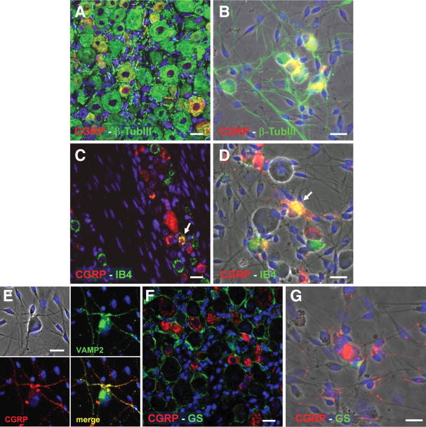
Figure 4.
Trigeminal neurons selectively express CGRP both ex vivo and in vitro. A, B, Double immunofluorescence staining of the CGRP peptide (red) and of the neuronal marker β-tubIII (green) in intact trigeminal ganglia (A) and in primary mixed trigeminal cultures after 48 h in vitro (B). C, D, Double immunofluorescence staining of the CGRP peptide (red) and FITC-conjugated IB4 (green), taken here as a marker of sensory neurons, in intact trigeminal ganglia (C) and in primary mixed trigeminal cultures after 48 h in vitro (D). Arrows indicate colocalization. E, Specific localization of CGRP (red) to VAMP2-positive axons terminals (green) in primary trigeminal cultures. F, G, CGRP (red) never colocalizes with the SGC marker GS (green). In all pictures, nuclei were labeled with the Hoechst 33258 dye (blue). Scale bars, 20 μm.

