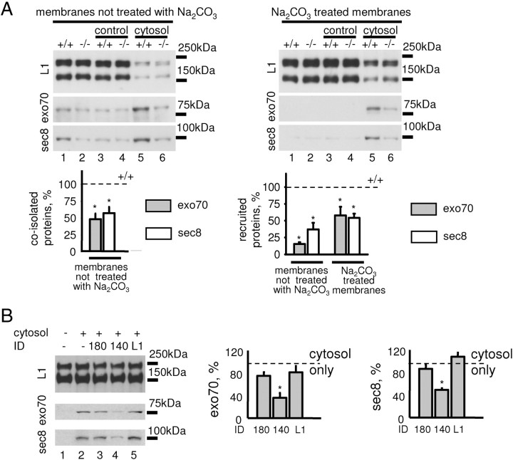
Figure 4.
NCAM recruits the exocyst complex to growth cone membranes. A, Recruitment of exo70 and sec8 from NCAM+/+ cytosol to NCAM+/+ and NCAM−/− growth cone membranes analyzed by Western blot with exo70 and sec8 antibodies. Control membranes were incubated with the buffer used for cytosol preparation. Labeling for L1 served as a loading control. Either untreated growth cone membranes (left) or growth cone membranes treated with alkali to strip peripheral proteins (right) were used. Note that levels of exo70 and sec8 coisolated with growth cone membranes are reduced in NCAM−/− growth cones (membranes not treated with Na2CO3, lanes 1 and 2). Note more efficient recruitment of exo70 and sec8 from the cytosol to NCAM+/+ (lane 5) versus NCAM−/− (lane 6) growth cone membranes either treated or not treated with Na2CO3. Graphs show mean + SEM optical densities in NCAM−/− probes normalized to NCAM+/+ levels set to 100% (dashed lines) from three experiments. *p < 0.05 (paired t test, compared to +/+ probes). B, Recruitment of exo70 and sec8 to NCAM+/+ growth cone membranes analyzed as in A. When indicated, the cytosol was preincubated with 180ID, 140ID, or L1ID. Note that recruitment is inhibited in the presence of 140ID (lane 4). Graphs show mean + SEM optical densities in 180ID-, 140ID-, and L1ID-treated probes normalized to the levels obtained with the cytosol only set to 100% (dashed line) from three experiments. *p < 0.05 (paired t test, compared to cytosol-only probes).

