
Figure 1.
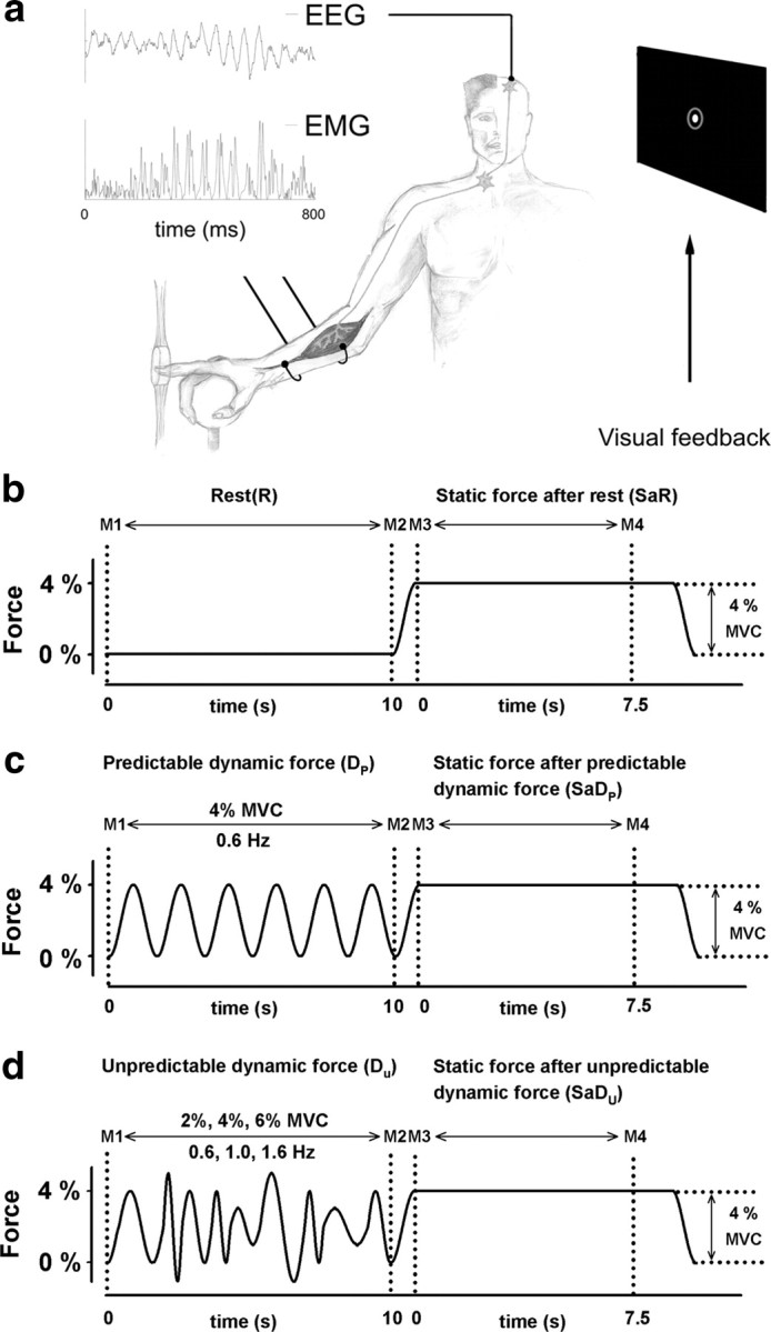
a, High-resolution EEG (52 scalp positions) and the EMG of the flexor digitorum superficialis muscle were recorded while the subject compensated the force profile of a manipulandum with the right index finger. Visual feedback about the position of the right index finger (white spot within the gray circle) was displayed on a monitor placed 60 cm in front of the subject and parallel to the face. EEG and EMG traces are shown during a period of high corticomuscular beta-range coherence in condition II. Simplified depiction of pyramidal tract, α-motoneuron, and muscle. b, Force profile generated by the manipulandum during one force trial of condition I. Condition I was made up of a rest period (R) in between the markers M1 and M2, and a static force period (SaR), in between the markers M3 and M4. c, Condition II consisted of a periodically modulated dynamic force period (Dp), in between the markers M1 and M2, and a static force period (SaDp), in between the markers M3 and M4. d, Condition III was made up of an unpredictable dynamic force period (Du), in between the markers M1 and M2, and a static force period (SaDu), in between the markers M3 and M4.