
Figure 6.
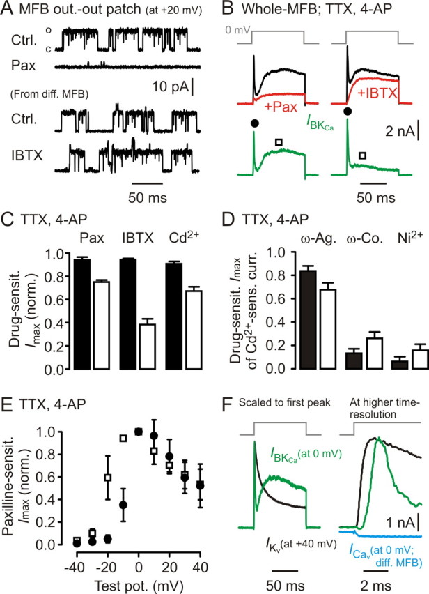
Two populations of presynaptic BKCa channels can be distinguished electrophysiologically at MFBs. A, Outside-out patches drawn from MFBs (steady state at +20 mV) exhibit large single-channel openings that are sensitive to Pax but not to IBTX. Ctrl, Control. B, Long depolarizing voltage steps to 0 mV in the whole-cell recording configuration reveal Pax- and IBTX-sensitive BKCa channel-mediated currents (green) composed of two kinetically distinct components. C, Summary of Pax (n = 4), IBTX (n = 7), and cadmium (Cd2+, n = 7) sensitivity for recordings as shown in B (filled bars, first, fast activating/fast inactivating component; open bars, second, slowly activating/persistent component; see also filled dots and open squares in B). D, ω-Agatoxin, ω-conotoxin, and Ni2+ sensitivity (each n = 6) of currents as recorded in B (bar code as in C). E, Voltage dependence of first, fast activating/fast inactivating component (open squares), and of second, slowly activating/persistent component (filled circles; n = 3). F, Left, Comparison of example Kv-mediated current kinetics (black trace) and BKCa-mediated current kinetics (green trace: Cd2+-sensitive current); traces scaled to the same peak amplitude. Note the extremely fast inactivation of IBKCa. Right, Same traces at higher temporal resolution. Blue trace represents example Cav-mediated current recorded in a separate MFB. Vertical scale bar applies to BKCa- and Cav-mediated currents.