Figure 5.
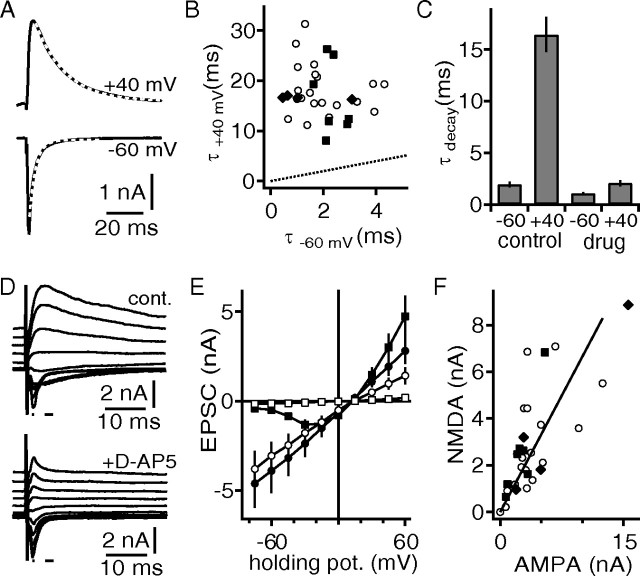
Excitation is mediated by AMPAR and NMDAR currents in DNLL neurons from juvenile gerbils. A, Synaptically evoked EPSCs recorded at −60 and +40 mV holding potential. The white dotted lines indicate an exponential fit to the decay of the EPSCs. B, Decay time constants recorded at −60 and +40 mV. Shown are black squares and diamonds from cells in which d-AP5 or CPP, respectively, was subsequently applied. The black dotted line equals unity. C, Effect of blocking NMDAR currents with either d-AP5 or CPP on the decay time constants at different holding potentials. D, Synaptically evoked EPSC at different holding potentials before (top) and after (bottom) the application of d-AP5. The small lines indicate the region that was used to estimate the AMPAR and NMDAR currents. E, Synaptically evoked EPSC size as a function of holding potential. The circles indicate AMPAR, and the squares indicate NMDAR currents before (black) and after (open) application of d-AP5. F, Correlation of the absolute size of the NMDAR (holding potential, +45 mV) and AMPAR (holding potential, −60 mV) peak currents. The symbol code is as in B. The solid line represents a linear regression.

