
Figure 9.
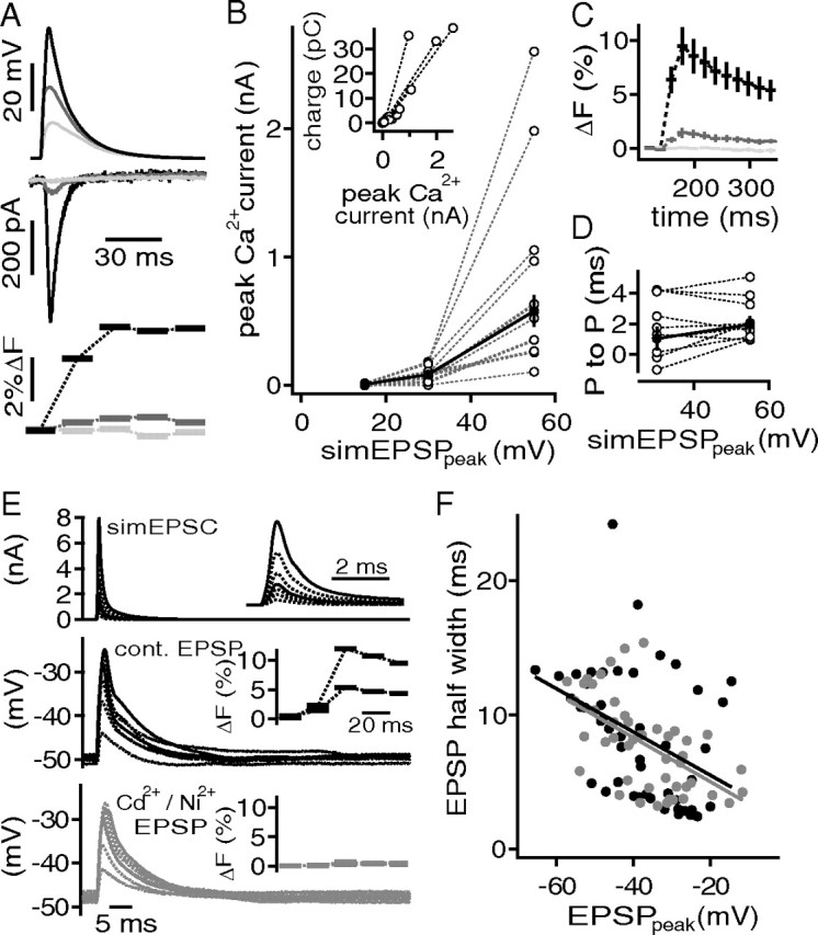
Ca2+ currents do not enhance spatial EPSP amplification. A, simEPSP waveforms (top) elicited pharmacologically isolated, and P/x corrected Ca2+ currents (middle) that were accompanied by a transient increase of flou-4 fluorescence (bottom). B, Ca2+ current as a function of the simEPSP peak. The inset shows charge of the Ca2+ current against peak amplitude of the Ca2+ current. C, Average fluorescence transient evoked with simEPSPs of 55 mV (black), 30 mV (gray), and 15 mV (light gray) amplitude. D, Time between the peak of the simEPSP and the Ca2+ current (P to P) for simEPSP amplitudes of 30 and 55 mV. E, Injection of different scaled simEPSCs (top) to evoke EPSP like deflections in control conditions (middle) and during blockade of Ca2+ channels (bottom). The insets show the magnified simEPSCs (top) and the fluo-4 transients (middle, bottom) that correspond to the EPSP traces shown in solid lines. F, EPSP half-width as a function of the absolute EPSP amplitude. The black symbols represent control conditions, and the gray symbols, during the application of Ni2+ and Cd2+. The solid lines are regression fits shown in corresponding colors.