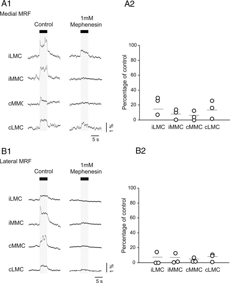
Figure 2.
Mephenesin strongly reduces the magnitude of the MRF-evoked responses in L2 MNs. A1, B1, Responses evoked during electrical stimulation of the medial MRF and lateral MRF with a 5 s train at 10 Hz (2 T) in the MNs of the iLMC, iMMC, cMMC, and cLMC before (left) and after (right) application of mephenesin to a split-bath compartment containing the cervico-thoraco-lumbar region of the spinal cord. Because individual labeled MNs are easily visualized through the ventral white matter, all optical recordings from MNs were performed in intact brainstem–spinal cord preparations (Szokol et al., 2008; Szokol and Perreault, 2009). A2, B2, Corresponding graphs showing the magnitudes of the responses during mephenesin application, normalized to the control response. Each point shows the average response in a single preparation and the horizontal lines indicate the grand mean.

