
Figure 4.
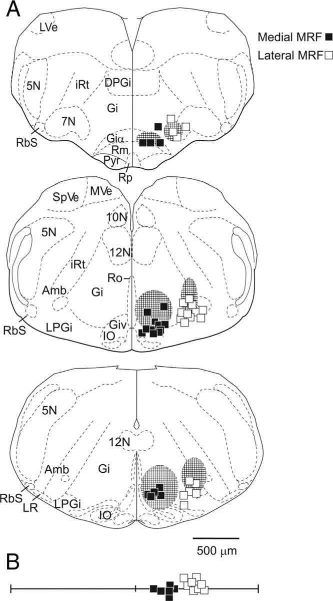
Locations of effective stimulation sites in the MRF. A, Stimulation sites recovered from transverse brainstem sections and plotted directly on standardized sections from a P0 mouse (adapted and modified from Paxinos et al., 2007) at three different anteroposterior levels. In all of these experiments, the locations of the stimulation sites were confirmed histologically from electrolytic lesions and DiI tracks left by the electrode trajectory (see Materials and Methods). The effective sites in the medial and lateral MRF regions are plotted as filled and unfilled squares, respectively. Cross-hatched areas indicate the regions that were previously found to be effective in evoking responses in the MMC and LMC (Szokol et al., 2008). 5N, Spinal trigeminal nucleus; 7N, facial nucleus; 10N, dorsal motor nucleus of the vagus; 12N, hypoglossal nucleus; Amb, ambiguus nucleus; DPGi, dorsal paragigantocellularis nucleus; Gi, gigantocellularis reticular nucleus; Giα, pars α; Giv, pars ventralis; IO, inferior olive nucleus; LPGi, lateral paragigantocellularis nucleus; LVe, lateral vestibular nucleus; LR, lateral reticular nucleus; iRt, intermediate reticular nucleus; MVe, medial vestibular nucleus; Pyr, pyramidal tract; RbS, rubrospinal tract; Rm, raphe nucleus magnus; Ro, raphe nucleus obscurus; Rp, raphe nucleus pallidus; SpVe, spinal vestibular nucleus. B, Additional effective sites for which electrolytic lesions were ambiguous or missing but mediolateral positions could be obtained from photographs of the electrodes positions during experiments. Scale bar: A, B, 500 μm.